Monday, October 4, 2010
Saturday, July 18, 2009
Scanning Electron Microscopy
So...I finally finished summer school a couple of weeks ago. I took Organic Chemistry + Lab and it was amazing! Anyways, I am finally getting around to posting some of the work I do with my research team at Appalachian State. These following images are pretty much the first images I came up with when I was learning how to operate the equipment last fall. These are all images of a common house fly, Musca dominica.
 This image shows the multiple hexagonal "ommatidia" that make up Musca dominica's compact eye. What is visible is the actual cornea of the ommatidia. Scanned at nearly 3000x magnification.
 This image is showing the area between the head of the fly and it's thorax. Quadrant 2 shows the proboscis of the head.
Friday, February 27, 2009
My Time at Appalachian
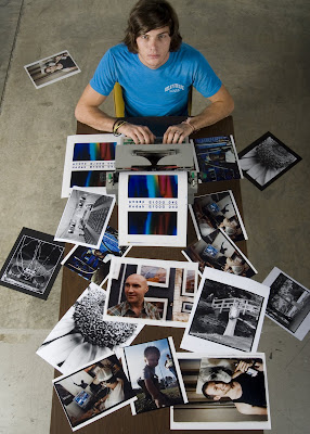
I have not done a lot of photography since I left RCC, but I have definitely kept busy. I am apart of research cluster in the microscopy facility here at Appalachian, and below are some samples of images that I have taken with the different equipment up here:
 This is an image of BPAE (Bovine Pulmonary Artery Epithelial) taken on a confocal microscope. The confocal microscope uses lasers from different wavelengths to excite particular dyes. These dyes can be "injected" into certain anatomy, and after exciting them with a specific wavelength of light, they become "excited" and visible. Here,the BPAE cells were labeled with MitoTracker® Red and BODIPY® FL phallacidin to show mitochondria and actin filaments.
The Following images are from a compound microscope. These were used in a research presentation for my microscopy cluster at the SNCURCS (State of North Carolina Undergraduate Research and Creativity Symposium)in November of last year. The final poster presentation is shown as the last image.
 Clinorchis Sinensis (Human Blood Fluke)
Anterior sucker and oral cavity
 Trypanosoma Gambiense (40x magnification)
This little parasite causes African Sleeping Sickness and is contracted from the bite of the Tsetse Fly
 Oral Smear (40x)
These are cheek epithelial cells covered in bacteria, with a visible bacteria strain
Wednesday, July 23, 2008
Fundus Photography - PDR & Asteroid Hyalosis

This is a composite of two fundus images taken of THE SAME patient, a 39 yr. old African female. The left image is ocular dexter and the right image is ocular sinister. I just thought the differences between the two were pretty interesting.
The right eye(left image) shows proliferative diabetic retinopathy(PDR), while the left eye(right image) shows extreme asteroid hyalosis.
ZEISS Fundus Camera
Fundus Photography - Optic Disc

This is just me messing around during free time at work. This is a triple composite. First, the background is a composite of all of the disc photos I have shot since working at the WFUBMC Eye Center. Then I put a fundus image of an optic disc with papilledema in front of that and masked some areas.
This is actually super corny, and I am usually against stuff like this. But whatever.
All shot on ZEISS Fundus Camera
Fundus Photography - Malignant Melanoma
 Sorry for the delay in posting, it has been very busy at my internship, and I have been trying to sort everything out for graduation as well.
 Anyways, here is an image of a malignant melanoma attached to the retina of the right eye in a 20 yr. old Caucasian male. Another was found in the left eye, but was about 1/20th the size of this one. If you look close enough you can really see the detail within this tumor. The retina itself is out of focus, but only because I wanted focus on the tumor.
ZEISS Fundus Camera
Wednesday, June 25, 2008
Slit Lamp - Keratic Precipitates

This is a photograph of Keratic Precipitates located on the inner endothelial layer of the cornea. This patient had a bad reaction to silicon oil used after a cataract removal and these precipitates formed.
KERATIC PRECIPITATES: Inflammatory cells and white blood cells from the iris and ciliary body that enter the aqueous and adhere to the innermost corneal surface (endothelium).
What is seen where the lens should be is not actually a cataract, because that was removed, it is actually part of the reaction to the silicon oil building up anterior the lens.
Wednesday, June 18, 2008
Slit Lamp - Post Traumatic Scleritis
 Another Slit-lamp image taken of a patient with Scleritis after trauma. Most patients we shoot on the Slit-lamp are photo-phobic (extremely sensitive to light) because of their anterior pathology, and it is not often you have a cooperative patient. They always pull away after every shot and complain. For this patient, we had to have three people in the room; myself, taking the photos, and two technicians forcing the patients head forward and pulling the eyelids open.
Also, sometimes the doctors will come in the camera room and want to watch, which was also the case for this patient, who was to immediately go into surgery after I finished the photographs. No pressure.
SCLERITIS: Inflammation of the Sclera
Slit-Lamp Camera:
Nikon D100's
Slit Lamp - Pterygium
Below is an image of what is called Pterygium. The "P" is silent.
PTERYGIUM: An abnormal, wedge-shaped growth on the bulbar conjuctiva, often related to sun irritation.
PTERYGIUM: An abnormal, wedge-shaped growth on the bulbar conjuctiva, often related to sun irritation.

Slit-Lamp Camera:
Nikon D100's
Tuesday, June 10, 2008
Macular Nevus

This is a nevus located beneath the macula in the choroid layer of the eye.
NEVUS: (Mole) Small, flat, usually pigmented benign tumor made up of specific cells called nevus cells; found in skin and eye tissues.
Zeiss fundus camera
Monday, June 9, 2008
The Operating Room: DSEK
 This is a shot taken in the operating room during a DSEK procedure.
 DSEK:
Descemet's Stripping Endothelial Keratoplasty.
Descemet's Stripping Endothelial Keratoplasty.
This procedure is done to replace and repair the tissue of the cornea. Instead of a corneal transplant that damages many endothelial cells and takes a long time to recover from, this procedure takes a donor cornea endothelial layer and places it through a small incision, allowing less recovery time as well as a lot less damage to the corneal cells. The following image shows the step by step process, from the donor cornea in the first image, to the placement and unfolding of the donor corneal layer, to the injection of an air bubble to hold the graft in place for proper healing position.
*This same patient had a cataract removed and an IOL (Intra-Ocular Lens) inserted just minutes before the DSEK procedure.
 Nikon D70x
ISO:200
105mm micro w/ PN-ll extension tube
F/16 w/ ringflash 1/2 power
Cataract Composite

This image is a composite of 4 images taken on the slit-lamp camera. Each separate image is called a red-reflex image, because you are lighting the posterior of the eye, and focusing on the anterior lens, so the cataract can be viewed somewhat like a silhouette. So two of the photos were at the nearest plane of sharp focus, while the other two were at the farthest plane of sharp focus. I used Photoshop to mask them together to show the full depth of the cataract.
Definition of Cataract: Opacity or cloudiness on the crystalline lens, which may prevent a clear image from forming on the retina.
Slit lamp: Nikon D100's
ISO: 200
F/22 w/ constant flash
Definition of Cataract: Opacity or cloudiness on the crystalline lens, which may prevent a clear image from forming on the retina.
Slit lamp: Nikon D100's
ISO: 200
F/22 w/ constant flash
Friday, June 6, 2008
Pyogenic Granuloma
This is an external shot of a pyogenic Granuloma. A breakdown of what exactly this means:
Pyogenic: Producing Pus.
Pyogenic: Producing Pus.
Granuloma: Dense collection of cells consisting of various inflammatory types.
THEREFORE: This is a growth of epitheliod (skin) cells, filled with pus.
Nikon D70xTHEREFORE: This is a growth of epitheliod (skin) cells, filled with pus.

ISO:200
60mm macro
F/22 @ 1/80
Finally....
Oh blogger where art thou? It's been about three months since I have even looked at one of these bad boys. I decided today that I will begin posting again due to the completion of my biology classes as well as the completion of my college admissions stuff. So in other words, I'm bored.
Most of what I'll be posting from now on is likely to be relative to my internship at Wake Forest University Eye Center as an Ophthalmic Photographer. I am not going to go into depth describing what is in these images, but only simple explanations for easy understanding. I hope everyone enjoys them.
This first image is a fundus(the inside) image of my own eyes. These were taken by David Miller, a co-worker of mine and a graduate of RCC. The photos are in sequence, right eye, left eye. This is also referred to as OD(Ocular Dexter), OS(Ocular Sinister).

Tuesday, February 26, 2008
Subscribe to:
Posts (Atom)