
 This is an image of BPAE (Bovine Pulmonary Artery Epithelial) taken on a confocal microscope. The confocal microscope uses lasers from different wavelengths to excite particular dyes. These dyes can be "injected" into certain anatomy, and after exciting them with a specific wavelength of light, they become "excited" and visible. Here,the BPAE cells were labeled with MitoTracker® Red and BODIPY® FL phallacidin to show mitochondria and actin filaments.
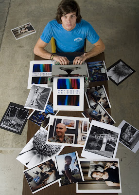
The Following images are from a compound microscope. These were used in a research presentation for my microscopy cluster at the SNCURCS (State of North Carolina Undergraduate Research and Creativity Symposium)in November of last year. The final poster presentation is shown as the last image.
 Clinorchis Sinensis (Human Blood Fluke)
Anterior sucker and oral cavity
 Trypanosoma Gambiense (40x magnification)
This little parasite causes African Sleeping Sickness and is contracted from the bite of the Tsetse Fly
 Oral Smear (40x)
These are cheek epithelial cells covered in bacteria, with a visible bacteria strain
3 comments:
even though i had no idea what half of those words are in your description...i gotta say, this is really effin' cool.
This is a nice blog....can i get a side of YOOOUUNN!!!
AWESOME!!
I'm mad jealous! Ive seen a few scopes in the Duke pathology labs; looks like your having a blast!!
Post a Comment